Topcon’s CV-5000 Automated Phoropter
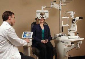 The CV 5000 automated phoropter offers all the latest features in automated refraction. It features a high-speed lens chamber, small optical head and several controllers.
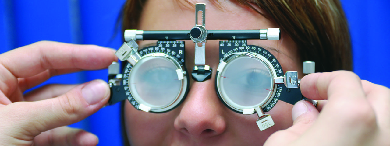
Topcon’s CV-5000 Vision Tester automatically imports data from pre-test instruments, making the information gathering and processing more streamlined. The unique color-coded cross cylinder makes it an easy-to-use system as well. Along with a digital eye chart, the procedure documents results in an electronic medical record for the clinic and streamlines the examination. The optometrist can control the lenses and letters used on the digital chart electronically with a keypad. The digital eye chart ensures good contrast and clear images to improve the accuracy and reliability of the eye exam.
The Ocular Response Analyzer (ORA) is the only instrument capable of measuring Corneal Hysteresis (CH); an indication of the biomechanical properties of the cornea. This information is different from thickness or topography, which are geometrical attributes of the cornea. Corneal Hysteresis represents a tissue property, which provides more comprehensive information about ocular biomechanics.
The Corneal Hysteresis measurement has been shown to be consistently associated with, or predictive of, rate of glaucoma visual field progression. In addition, corneal biomechanics are the primary influence on tonometer accuracy. Ocular Response Analyzer’s ability to measure Corneal Hysteresis enables the device to provide corneal compensated IOP, called IOPcc, which has been proven to be less influenced by corneal properties than Goldmann or other methods of tonometry.
We use advanced digital imaging technology to aid in the assessment of your eyes. Like most diseases early detection is key to a good prognosis and eye diseases are no different if detected early then most can be treated to prevent loss or slow the loss of vision. Images are stored electronically and electronically linked to your patient record. This gives us measurable and comparable data to monitor and gauge the effectiveness of treatment. Most eye conditions, such as glaucoma, diabetic retinopathy and macular degeneration are diagnosed by detecting changes over time.
CARL ZEISS Humphrey 740i Field Analyzer
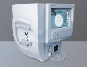 The CARL ZEISS Humphrey 740i Field Analyzer, is a tool for measuring the human visual field, it is used by optometrists, orthoptists and ophthalmologists, particularly for detecting monocular visual field.
The results of the CARL ZEISS Humphrey 740i Field Analyzer identify the type of vision defect. Therefore, it provides information regarding the location of any disease processes or lesion(s) throughout the visual pathway. This guides and contributes to the diagnosis of the condition affecting the patient’s vision. These results are stored and used for monitoring the progression of vision loss and the patient’s condition.
Topcon Maestro 2
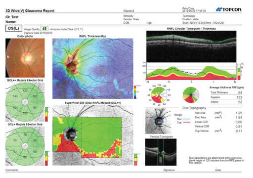
The Maestro2 can capture a 12mmx9mm widefield OCT scan, encompassing both the macula and optic disc. Ideal for an annual eye exam, the scan reduces patient testing time. It provides thickness and reference data for the retina, RNFL and ganglion cell layers together with a Glaucoma report which includes disc topography.
Neurolens
Neurolens glasses offer a groundbreaking solution to alleviate headaches, eye strain, and discomfort in the neck and shoulders by addressing eye misalignment. If you’re plagued by chronic headaches due to digital eye strain or close-up work, neurolenses may be the answer you’ve been seeking. These innovative prescription lenses are uniquely crafted with a contoured prism that effectively realigns your eyes at varying distances, tackling issues related to trigeminal nerve pressure.
The key to neurolenses’ success lies in their specialized measurement diagnostic tool, designed to identify potential eye misalignment and its extent. In a brief, painless examination lasting under three minutes, the neurolens measurement device assesses the gap between where your eyes should be and their actual position, thus determining the degree of synchronization and alignment required. The result is a tailored contoured prism prescription that can significantly improve your visual comfort and alleviate the discomfort associated with eye strain and misalignment. Neurolens glasses are a promising solution for those in need of relief from the debilitating effects of eye-related issues.
OPTOS Daytona Plus (Optomap Retinal Exam)
Annual eye exams are vital to maintaining your vision and overall health. We offer the optomap® Retinal Exam as an important part of our eye exams. The optomap® Retinal Exam produces an image that is as unique as you fingerprint and provides us with a wide view to look at the health of your retina. The retina is the part of your eye that captures the image of what you are looking at, similar to film in a camera.
Many eye problems can develop without you knowing. You may not even notice any change in your sight. But, diseases such as macular degeneration, glaucoma, retinal tears or detachments, and other health problems such as diabetes and high blood pressure can be seen with a thorough exam of the retina. We've recently received a brand new Optos Daytona Plus imaging device that incorporates the newest hardware and software technology that allows us to see more, discover more and effectively treat and manage any ocular disease.
An Optos Optomap Retinal Exam provides:
- A scan to show a healthy eye or detect disease.
- A view of the retina, giving your doctor a more detailed view than he/she can get by other means.
- The opportunity for you to view and discuss the optomap® image of your eye with your doctor at the time of your exam.
- A permanent record for your file, which allows us to view your images each year to look for changes.
The Optos Optomap Retinal Exam is fast, easy, and comfortable for all ages. To have the exam, you simply look into the device one eye at a time and you will see a comfortable flash of light to let you know the image of your retina has been taken. The Optos Optomap image is shown immediately on a computer screen so we can review it with you.